When it comes to non-invasive ventilation the field has become a little more crowded in recent years at least in our institution. In the recent past if one decided to extubate an ELGAN the biggest decision was what CPAP pressure to use. These days we have the option of high frequency nasal ventilation (nHFOV) or non-invasive positive pressure ventilation (NIPPV) to choose from as additional options. Both of these modalities have their uses and I have written about nHFOV before as in Nasal High Frequency Oscillatory Ventilation For Preventing Intubation. On this post though I want to look at NIPPV which has actually been around longer as a modality. The gist of this mode is that one chooses a delta P, peep, Ti and rate much like you would on a conventional ventilator. When ventilating through a nasal interface the device provides ventilation although it is questionable I suppose how much of that is alveolar ventilation. The study we are going to talk about here caught my eye as the information gleaned from it gives me at least an idea of how this mode may work to help prevent reintubations.
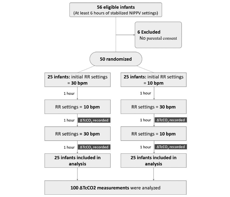
In this study each patient served as their own control and alternated between either a start of a rate of 10 BPM or a rate of 30 BPM as shown in the following diagram. The infants were all between 24 +0 and 32 +6 weeks gestation to be included in the study. Delivery of NIPPV was through the Leoni Ventilator using RAM cannulae and importantly the mode was non-synchronized. Each infant needed to be stable on their settings for at least 6 hours before being included. The authors hypothesis was that rate matters to clear carbon dioxide. To monitor CO2 levels they used transcutaneous CO2 measurements to allow for continuous measurement over each hour of the study. Given this belief, there was safety built into the protocol such that patients were excluded if on the set rate of 10 bpm the tcCO2-related pCO2 was <40 mmHg, or on NIPPV if the set rate of 30 bpm had a tcCO2-related-pCO2 is 60 mmHg, In other words, if rate matters and your tcCO2 was already less than 40 on a low rate then it would not be safe to blow off more CO2 and vice versa with high CO2 and low rates. To ensure that only rate affected the results “during the 3 h of the study no changes in PIP, PEEP or FiO2 were allowed with the following exceptions: if spO2 was <90% or >95% for more than 20 s, an increase or decrease in FiO2 were allowed to keep spO2 90–94%, and were documented”.
So does rate matter?
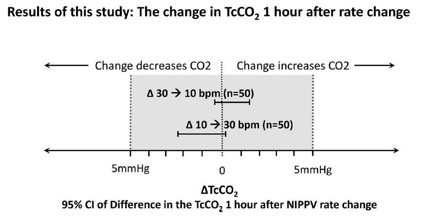
It turns out the authors found no difference in CO2 levels based on rate changes alone.
This of course is contrary to what the authors expected to find. The question is why this might be. What follows now is just speculation on my part but given the finding of no difference I can offer a few thoughts. The first is that NIPPV does not involve a distal delivery of gas like the situation of an endotracheal tube near the carina. With an endotracheal tube in place the delta P or pressure above the set peep is delivered to the gas exchanging areas of the lung. With NIPPV you are delivering the pressure at the nose and therefore there is a fair amount of dead space in between the exit of the gas into the baby and the lung. Might you just be really ventilating dead space for the most part?
Secondly, depending on the fit of the mask or the degree that the mouth was open how do we know how much of the non-invasive ventilation reached the infant? Lastly, in our own centre we have not been impressed with the RAM cannulae as we have found that whether the prongs are in or out of the nose the pressure being detected as being delivered seems to stay the same at least as the ventilator sees it. If the prongs were not in the nose properly and the atmosphere was being ventilated would one really know that the pressures weren’t really getting into the nose?
Lastly, the Leoni ventilator is not capable of delivering synchronized NIPPV. Now that there is the availability of synchronization on ventilators such as on the Puritan Bennett 980 ventilator it would be interesting to see the same study done again. If you are delivering non-synchronized breaths which are not in sync with the patient should we expect a change in CO2? What if half the breaths for example by chance are delivered on exhalation? Not much effect on CO2 I would think.
I am not saying that rate doesn’t matter at all but I suppose I am saying within the context of this study it doesn’t matter to CO2. My best guess as to how NIPPV works to prevent reintubation may be secondary to two things. The first would be by irritating the baby with the puffs of delta P. Think of it like intermittent stimulation. The second possibility is that the same puffs of air help keep the pharynx open and minimizes the obstructive portion of apnea of prematurity. Whatever the reason NIPPV appears to work to prevent reintubation in some infants!
I have no doubt the group here will look at the effect of delta P on CO2 soon enough and I wonder if we will see much difference there either. It also will be important to look at the effect of rate in a synchronized fashion! Time will tell.
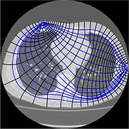
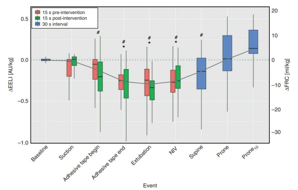
Extubation is a regular occurrence in the NICU. We do our best to predict who will succeed and who will fail but it isn’t always easy to figure out who they are in advance. We use techniques such as looking at oxygenation histograms and using thresholds for PIP, PEEP or MAP but in the end sometimes it works and other times it doesn’t. In an effort to improve on intubation success, some creative researchers in Switzerland employed a technique called end-expiratory lung impedance or EELI to measure lung volume before, during and after the extubation process. The use of EELI is based on the impendance of the lung changing with the distribution of tissue and air and by placing electrodes one can generate a cross sectional volume that has been shown in neonates to be representative of total lung volume. The EELI technique creates an image like this which is use to generate the estimate of lung volume.
The researchers in this study were seeking to do a quality improvement project and use EELI to estimate lung volume at different time points in an extubation. The time points were all 30 seconds including, immediately before first handling of the infant (baseline), tracheal suctioning (suction), start and end of adhesive tape removal (adhesive tape begin and adhesive tape end), pulling the endotracheal tube (extubation), initiation of non-invasive ventilation (NIV), immediately before and after turning the infant to prone position (supine and prone, respectively), and 10 min after turning to prone position (prone10). As per unit policy all babies were ventilated with Draeger VN500 ventilators and if <28 weeks went on to NIPPV when extubated or if 28 weeks or more straight CPAP. The purpose of this quality initiative was to determine using EELI at what point in the extubation process infants might be losing lung volume and then based on the information see if they could ultimately use this to improve the chances of successful extubation in the future.
What makes this study interesting is that the infants were found to lose volume but at a time when I would not have expected it.
The Reveal
Below is a graphical depiction of EELI and estimates of FRC during the different time points. The changes in electrical impedance by EELI were converted on the right Y axis to an FRC in mL/kg.
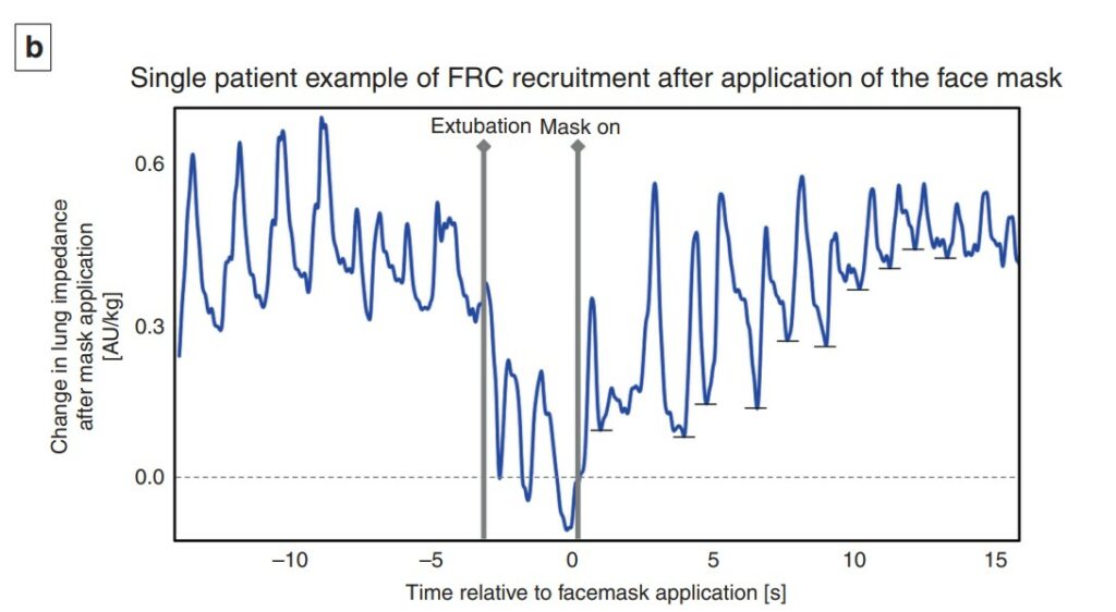
What is surprising at least to me here is the loss of volume occurs not with extubation but rather when the tape removal process happens. With the placement of the prongs on the infant at extubation the FRC gradually rises and recovery occurs. Moreover as shown in the 12 patients included in this study, the recovery once non-invasive ventilation is provided is quite rapid and evident within 1-2 breaths.
A couple other things to note. The loss of FRC during tape removal was about 10 mL/kg and if typical FRC in a preterm infant is 20-25 mL/kg you can see the impact this would have on lung volume and reserve. As this was a small study it could not detect a threshold at which extubation would fail but one infant who developed a pneumothorax and required reintubation did not get back to their baseline FRC.
What is this signaling?
Yes this is a small study but it did look at about 3000 breaths so there is a fair amount of data to look at. What the paper demonstrates I think is that there is a vulnerable time during tape removal where likely due to the fact that we use uncuffed ETTs in neonatology it is possible for these infants to lose lung volume. It may be that as they strain and bear down the ventilator may not be as effective at delivering volume to them. Measures that might help during this time could be skin to skin care, breastmilk drops or scent, sucrose or a variety of other non-pharmacologic measures to keep them calm. This might help to minimize such volume loss. Secondly, knowing the significant risk of volume loss it underlines the importance of placing nasal prongs on as quickly as possible during the transition from invasive to non-invasive ventilation as recovery of lung volume is possible. It think it also suggests that if we are “peepaphobic” and use an insufficient amount of support at extubation these infants may be vulnerable to experience significant volume loss as well.
While EELI may not be perfect, this study is the first of its kind and may shed some light into why some infants fail after extubation. While usually I say less is more, I do wonder if in the case of extubation, this study gives some evidence to support starting with a higher PEEP than you think you need non-invasively and then backing off after one has successfully extubated. This may be the first study I have seen on this but I am certain it won’t be the last.
When I began my career in Neonatology we initially ventilated primarily with pressure limited time cycled modes of ventilation and only supported some of the breaths as in SIMV modes. With time and emerging research a movement to using set volumes came about and in many centres supporting every breath using an assist control mode or similar version. Although I don’t have access to it in my centre, ventilators such as the Avea also allow for automated FiO2 control in addition to having a choice of two different volume targeting modes. The difference between the modes is the subject of a study entitled Comparison of volume guarantee and volume-controlled ventilation both using closed loop inspired oxygen in preterm infants: a randomised crossover study (CLIO-VG study). I suppose it shouldn’t be a big surprise that as technology advances and we fine tune practice, different modes for volume targeting would arise.
What’s the difference?
Volume Controlled Ventilation (VCV) – flow based on a set volume and measurement of the PIP every 2 ms. Next breath is given the greater of PEEP+2 cm H2O or PIP-2 cmH2O. The ventilator at the end of each breath is able to determine if the baby is still demanding flow and continues providing flow but stops when inspiratory flow is <25% of peak set flow. There is constant inspiratory flow and peak volumes.
Volume Guarantee (VG)- inspiratory pressure is adjusted breath to breath. These breaths have a decelerating flow instead of a fixed flow as in VCV.
The Study
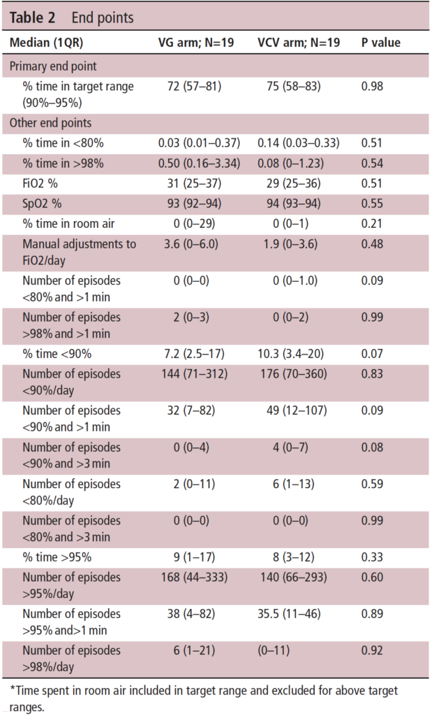
Using automated FiO2 control for both groups the study design was a crossover one. The concept was that better ventilation would help to keep O2 saturations more reliably in a target range of 90-95% and that one of these modes might be superior than the other in doing so. Infants in the study were born at 23+0 – 36+6 weeks and had to be intubated and on >21% FiO2 to be part of the study. Each group spent 12 hours in each arm with the starting mode randomly chosen before switching over to the other mode.
Based on a power calculation in which the authors selected looking for a 5% difference they determined they needed 19 patients in the study overall. The median GA of the infants was 25 weeks (IQR 24-28) with a BW of 685g.
The results demonstrate at the top of Table 2 that the primary outcome was no different at all. Basically whichever mode you choose will work just fine when used with automated FiO2 control to keep the saturations in the target range. If there is anything that the study suggests though is that the percentage of time below 90% may be worse with VCV than VG. You get this from looking at the table and looking at the secondary outcomes. A word of warning though that since the study is small (very small) it is really difficult to take too much stock in the secondary outcomes as the study wasn’t powered to detect such differences. One can’t help but wonder though if that trend might have become a one of significance if the numbers in the study were greater. Is there biological plausibility for this? Looking at the two modes, it would appear that VG by adjusting each breath based on the last expired tidal volume may be more agile. If you believe the hypothesis that tighter control of alveolar ventilation by delivering better ventilation is key to reducing time outside the target ranges then it makes some sense that this mode would be better.
On a personal note, I use only VG in my centre so I am pleased to see there is really no difference in the primary outcome but the trend in the secondary outcomes at least puts a slight smile on my face as well!
Who doesn’t love a good match up?! Supporting neonates in need of resuscitation after delivery has been the subject of many studies over the years. The movement has certainly been to non-invasive support with CPAP or NIPPV but some babies need some degree of support with PPV after delivery when they simply won’t breathe. Prior to intubation the rise of the t-piece resuscitator has meant that practitioners can set a PIP and PEEP and with only a finger press to deliver a tidal volume at set pressure and with the finger released provide CPAP through the same device. The only problem potentially with use of these devices is the imposed work of breathing (iWOB) which has been measured in other studies. Any device I have used has provided ventilation through a mask so imagine my surprise to come across a new device called rPAP using prongs from the original infant flow design. From the manufacturers website the company claims that their design used with either a mask or nasal prongs reduces iWOB by 92% compared to other comparable machines! Imagine my greater surprise to see a head to head RCT comparing this new device to standard t-piece resuscitators with a mask.
The intervention was completed with one of three outcomes were met.
Infant intubated
Stable and breathing on method of support after a minimum of 10 minutes of support.
At 30 minutes when respiratory support could continue as decided by the clinician without crossover allowed.
Looking at the appendices for the trial it appears that one could use either device to administer PPV or CPAP but the point of the trial was that the devices would be used to support the infants until one of the three above criteria were met. If the claims about reduced iWOB were true compared to other devices in use then one might expect to see a difference in the primary outcome of incidence of intubation or death within 30 minutes of birth.
In total there were 250 infants recruited with 127 assigned to the rPAP and the other 123 to t-piece resuscitation. The mean GA in the trial was 24.8 weeks and the baseline characteristics between groups were similar although the group randomized to the rPAP has more c-sections and more general anesthetic exposure compared to the t-piece group. Lastly, humidification of gases during resuscitation was similar between the two groups.
How Did They Compare?
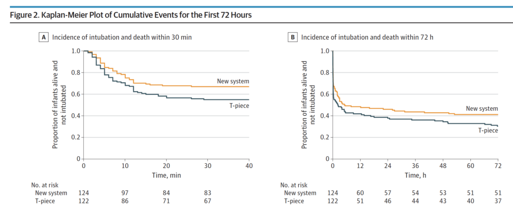
It just might be that the claims of decreased iWOB might have merit. In Figure 2 below the Kaplan-Meier curves show a difference favouring the rPAP device when looking at the primary outcome. This difference was significant with 41 of 124 infants (33.1%) in the rPAP group and in 55 of 122 infants (45.1%) in the T-piece group having the primary outcome of intubation or death within the first 30 minutes of life. Moreover when looking at the adjusted odds ratio it was still significant at 0.53; 95% CI, 0.30-0.94. The incidence of intubation and death in the first 72 hours although trending towards favouring the new system did not reach statistical significance.
Finally, none of the secondary outcomes reached statistical significance which included such things as death in the delivery room, use of surfactant, or PPV in the DR.
Does it make sense?
If you had asked me to tell you prior to the study whether resuscitation with nasal prongs vs a mask would be different I would have said a mask would be better due to less leak. Turns out based on this data that I would be wrong in that guess. A look at the website though for the rPAP device indicates that it can be used with a mask or nasal prongs. It would have been nice in the study presented here to have used a mask as a third arm with the rPAP device as it leaves me wondering a bit whether it was the interface that mattered more than the type of driver used? Maybe I am wrong and by using prongs it allows the infant to have less iWOB than with a mask over the mouth and nose? Could it be that it has more to do with that that the type of driver whether it is a traditional t-piece resuscitator or the new rPAP device? Regardless, I have a suspicion that these results will resonate with people. A posting of the abstract alone has garnered a lot of attention on twitter this week so clearly this is of interest.
I don’t think there is much fault to find in this study other than my question of why they didn’t choose to have a head to head comparison with masks as well but perhaps that is for another study. I imagine we will see this approach adopted in many centres around the world as they replace their traditional t-piece resuscitators in need of replacement. I also suspect there will be many that will want a larger study before adopting this strategy to look more closely with come faith in the results at secondary outcomes in particular having to do with safety.
One thing is for certain. There will be more studies to come!
Here in Winnipeg we don’t use t-piece resuscitators for any resuscitation. I did use them in my past position in Edmonton and I came to appreciate them for their ease of use. For the majority of infants, setting a PIP and a PEEP and then using your finger to occlude and release offers a relatively simple and less difficult approach to ventilation than using a self inflating or jackson-rees bag. I say the majority of infants, as most infants are not born from 22-32 weeks but the lion’s share are born at gestations older than that. The larger more mature infants have lungs that are much more forgiving to excessive ventilation. For the smallest of infants though questions have remained for some time around the volumes delivered to the fragile lung when a fixed pressure is used in the presence of moment to moment changes in compliance.
Measuring Tidal Volume in Intubated At Risk Infants
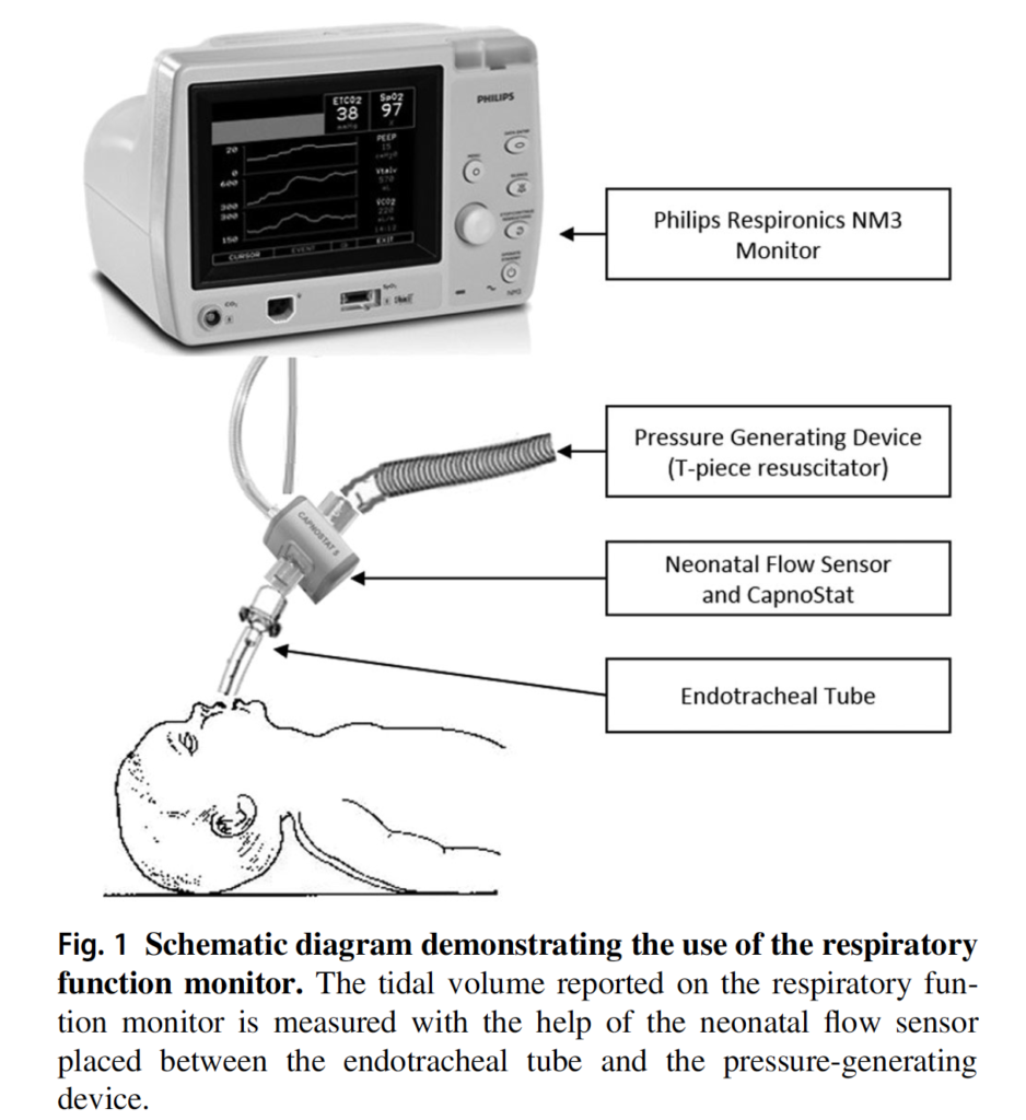
Vaidya R et al published Tidal volume measurements in the delivery room in preterm infants requiring positive pressure ventilation via endotracheal tube feasibility study in Journal of Perinatology. The prospective observational study looked at 10 infants born at < 32 weeks with a mean GA of 23.9(±1.5) weeks and mean BW 618.5(±155)g. A mean of 17.8 minutes of recordings were examined using the setup below and in total looked at 8175 individual breaths. All patients in the study were intubated with non-cuffed ETT but by only including intubated infants in the delivery room the issue of mask leak was avoided. As in many units the target Vt was 4-6 mL/kg. It wasn’t specified what criteria they use for setting initial pressures but the included patients had a mean PIP of 24.4±5 and PEEP of 5.9 ±2.4. Importantly, those providing ventilation with the t-piece resuscitator were blinded to the data on tidal volume measurements.
How Good Were They At Meeting Their Goal?
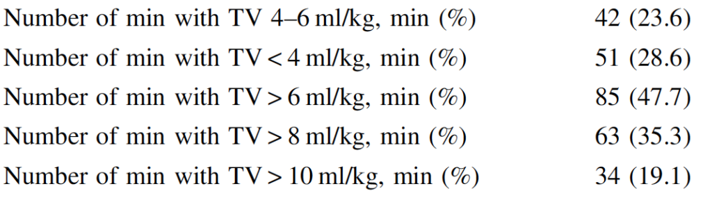
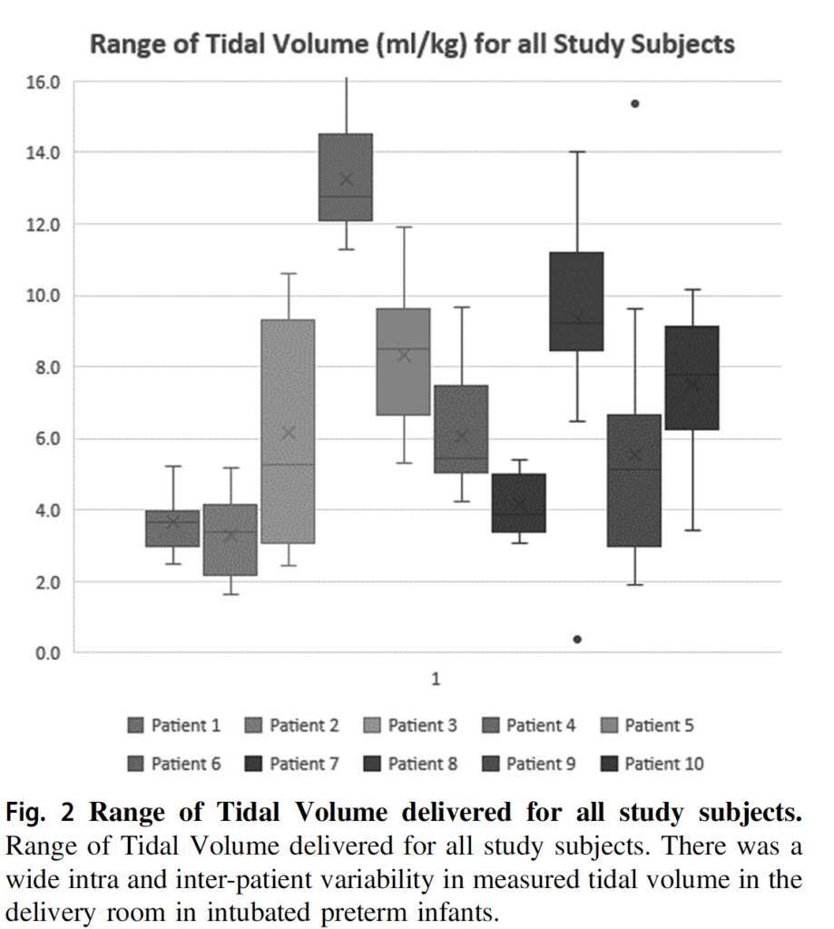
It turns out that they weren’t that great (I am not faulting them by the way) as it is a challenge to try and adjust pressures based on chest rise. We are not good at it at all. As shown in the figure below there was a wide range of volumes administered. In fact here is the breakdown. The goal Vt between 4-6 was only 25% of the time. In other words you are dealing with either a risk of atelectotrauma or volutrauma 75% of the time. It is worth noting that the neonatal flow sensor has a dead space of 1 mL. If that is the case and the infants on average were about 600g that is almost 2 mL/kg in non-ventilated space that this volume is going into. It doesn’t change the numbers that much if you factor that in but it does mean that some infants who were getting a measured 3 mL/kg were actually seeing under 2 mL/kg of lung ventilation. On the other hand those getting 7 mL/kg were actually seeing under 6 mL/kg so were in target. Bottom line though is that when using fixed pressure settings in the presence of changing compliance even if one is adjusting pressure in real time it is difficult to maintain stable volumes in target range. The authors also demonstrate in another graph that even in individual patients there is fluctuation as well.
Call to Action
I think this study is actually quite useful in confirming what I imagine many have always suspected. We just aren’t that great at assessing tidal volume when we watch the chest rise. As many have noted, the first 6 breaths at least in an animal model can damage the lungs. Imagine what excessive or low volumes can do to the lung over 18 minutes?!
What this study does is demonstrate especially in the smallest and most vulnerable infants that if ventilation is needed one should put the infant onto a volume guaranteed mode of ventilation ASAP. Ventilators should be in the resuscitation area as we have in our hospital and not have to be brought in should the baby be intubated. Hand bagging even with a t-piece resuscitator should be kept to a minimum. At risk is the development of BPD and knowing that even in experienced hands we just aren’t that good at delivering tidal volumes in a target range we need to strive to minimize the time that we expose our infants to such modalities. Ventilation isn’t always avoidable but when needed my advice is to control volume and allow pressures to fluctuate as resistance and compliance change. Especially after administration of surfactant the pulmonary mechanics are changing constantly and no matter how good you are you just won’t be able to keep pace. Let the ventilator do it!
Giving bronchodilators to preemies on a ventilator has certainly been tried before. The major issue to contend with is getting the drug to where it is supposed to be. Anyone reading who has a child with asthma knows that you should use an aerochamber when taking a puff to help with better distribution to the lung. Giving a puff or two without it largely ends up on the back of the throat. Similarly, giving puffs through an endotracheal tuberaises questions about how much of the medication winds up on the plastic tube rather than the smooth muscle of the airways where the medication is intended to be. This has been looked at in a cochrane review as well entitled Bronchodilators for the prevention and treatment of chronic lung disease in preterm infants
Can Albuterol Save The Day?
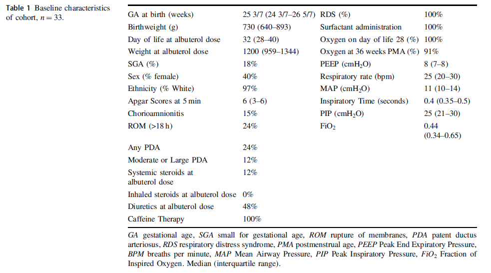
Albuterol is a beta agonist much like ventolin that can act on the smooth muscle of airways to achieve bronchodilation. Considering that preemies with immature lungs may have issues with both resistance and compliance, Raffay TM et al in their paper Response to first dose of inhaled albuterol in mechanically ventilated preterm infants chose to examine responsiveness in a group of 33 infants (all < 30 weeks at birth) to albuterol. Ideally, responsiveness would be done by pulmonary function testing but given that this was not possible in these infants they chose to examine other indicators of impact. After giving two puffs of 90 mcg of albeterol via a metered dose inhaler without an aerochamber the authors looked at changes in FiO2 as well as compliance and resistance measurements on the ventilator as a means of determining responsiveness. Ultimately, could they get drug into the distal airway in patients who were ventilated at about a month of age as shown in table 1 along with other baseline characteristics?
What makes this different than other studies I suppose is the use of the ventilator measurements and their use of histogram data on oxygen saturation to ascertain responsiveness to treatment. This was an observational study based on a secondary analysis of a previous study so we don’t have sham controls to compare to. Having said that by administering the medication and seeing what happens immediately afterwards it is possibile to get a sense of whether the drug had an effect.
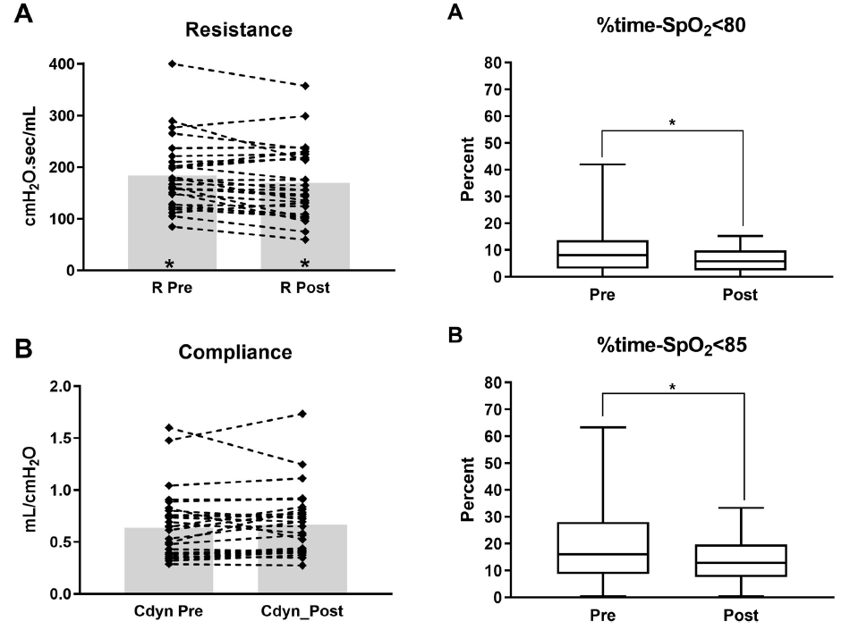
So What if Any Effect Did It Have?
From the figure in the paper the answer is some effect. Overall, post albuterol resistance for the 33 patients overall was found to decrease. Compliance and FiO2 (not shown in the graphs below) did not change though. What did change however was the percentage of time spent below 80 and 85% respectively comparing a 4 hour window pre and a 4 hour window post with respect to histograms from the patient monitor.
Putting it together
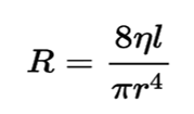
Ok so this isn’t a gold standard RCT looking at placebo treatments vs albuterol. It is hypothesis generating though as if resistance was decreased by albuterol one could expect improved delivery of O2 to the distal alveoli and therefore better oxygenation which is what is seen here. Should we be surprised that no difference in compliance is seen with albuterol therapy? I don’t think so as the effect of the drug is not on the distal alveoli and parenchyma but rather the more proximal branching airways. SInce airway resistance is governed by Poiseuille’s Law (you thought physics was over in high school?!) you can see that resistance (R) is directly proportional to the viscosity (n) and length (l) of the airway but inversely affected by the radius (r) to the 4th power. In other words if the radius of the airway after albuterol increases by 25% that effect is amplified to the 4th power in terms of reducing resistance.
I suppose I am buying what they are selling here but again the key is finding a method of getting the drug to deposit not in the trachea or proximal bronchi but to the lower airways. I can’t help but wonder if use of high frequency jet ventilation which carries flow down the centre of the airway might be a very effective way of getting such puffs further into the lung. Speculation of course but perhaps someone a little more creative than I can figure out how to do that and test deposition.
Should we use this routinely? Probably not as an everyday approach but it does make me wonder about those babies who are having a bad day so to speak. If one can glean from the ventilator that resistance has increased from one day to another might this be something worth trying? The authors found that the first treatment was effective but second and third not so much so to me this may just be a “hail mary” that is worth trying when nothing else seems to be working to reduce FiO2 in the presence of increased resistance.
If anyone is doing this routinely I would be curious in hearing your own experiences.