I spend a bit of time on social media and when I do I come across the argument that vaccines aren’t needed in pregnancy if you have already had COVID. The concept from the vaccine hesitant is based on the notion of trying to avoid any perceived risk of vaccination when the body is already making antibodies against the virus. The literature has been fairly scant on newborns in terms of protective antibodies and limited to case reports/series that I have shared from time to time on either twitter or facebook. As you might expect something might have changed as I am writing a piece on this topic again. The change is related to a recent paper entitled Titers of SARS CoV-2 antibodies in cord blood of neonates whose mothers contracted SARS CoV-2 (COVID-19) during pregnancy and in those whose mothers were vaccinated with mRNA to SARS CoV-2 during pregnancy by Kashani-Legumsky et al in J Perinatol.
Setting The Stage
Before getting in to what they did it is important to understand how the mRNA vaccines work as the antibodies that one can look at in mothers and babies are of two types. The mRNA vaccines instruct the body to make anti-bodies against the spike protein (S antibodies) which forms the basis of how the vaccine helps our bodies identify the virus and then destroy it. For those who have actually been exposed to the virus and are not vaccinated, they develop a second antibody to the nucelocapsid protein (N antibody) which is within the viral core so this type will only be present in people who have been infected with the virus and their immune systems have dealt with it on their own. This is an important distinction as it allows you to create pure samples of people who have had the virus as a true infection and those who have been vaccinated and finally those who are neither.
Comparing Three Groups
So the authors here decided to compare three groups of women. Eighty three cord blood samples were divided into three groups (from the paper quoted) based on IgG antibody titres.
Group 1 included 29 samples (37%) from women who were infected with SARS-CoV-2 during pregnancy. Twelve had RT-PCR confirmed Covid-19 infection: three were infected in the first trimester, three in the second trimester and six in the third trimester. The other 17 had no clinical signs of SARS-CoV-2 infection during pregnancy and had a positive serologic test on admission. None of the 17 women had active SARS-CoV-2 infection at the time of delivery. Group 2 included 29 samples (37%) from women who were vaccinated against SARS-CoV-2 in the 3rd trimester.
Group 3 included 21 women (34%) and served as controls.
Looking at antibody levels in Group 1&2, 100% were positive for S antibodies. Interestingly, in group 1, 4 women did not test positive for the N antibody (3 were asymptomatic and one infected in the 1st trimester). In group 3 none of the women tested positive for any antibodies confirming they were neither vaccinated or had the infection previously.
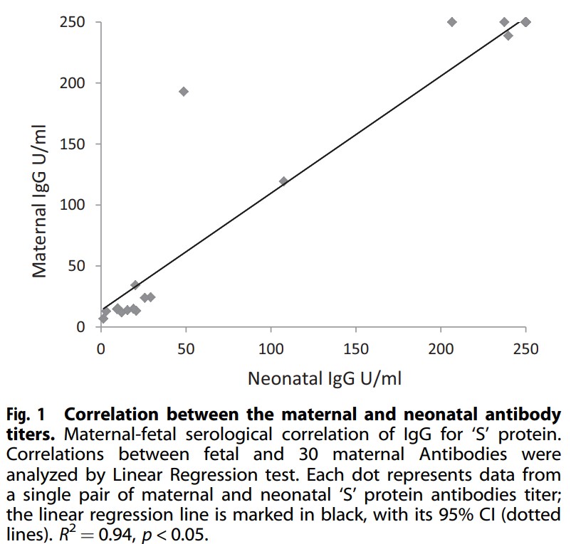
Looking at mean antibody S titres there was a significant difference found in that Group 1 had a mean of 83.7 U/mL vs 225.5 U/mL for the newborns whose mothers were vaccinated. Also notable was the relationship (not surprisingly between antibody levels in the mother at the time of delivery and newborn cord blood titres.
There was a linear correlation between the level in the mother and the level found in the newborn with higher levels presumably better for protecting the infant. Having said that, no infants in this study had neonatal COVID infection. Detractors would be quick to point out that this indicates it doesn’t matter if you get the vaccine since all babies were ok but remember although this may be the biggest study looking at antibodies in cord blood it remains a very small sample and neonatal infection although reported, remains a very rare occurrence.
The Other Side
If you have followed my coverage of the COVID saga from the start you would know that I am in favour of vaccination and in pregnancy as well. The results of this study are encouraging but we need to compare apples to apples. This study compared women who were vaccinated in the 3rd trimester to women who were infected at earlier time points and may have been sick or asymptomatic. The lower antibody levels found in group 1 could represent declining titres as the infection becomes more remote. What we also don’t know is what they antibody levels would have looked like in group 2 if the mothers were vaccinated in the 1st or 2nd trimester as this is now happening. Would the levels be similar? They just might be as the antibody levels do decline with time. We rely on memory cells to reactivate our antibody producing cells if the virus comes along again.
I am not saying this study is meaningless but be prepared if you quote this study for vaccine hesitant to point out that you are comparing recent vaccination to potentially mild cases or remote infections. What is clear and hopeful though is that your newborn is protected by antibodies you make in pregnancy from vaccination at very good levels and until we can vaccinate babies this is the greatest protection we can offer.
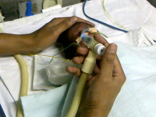
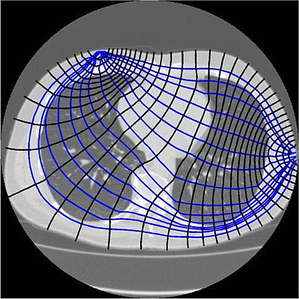
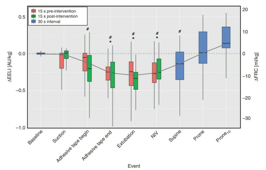
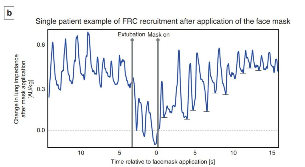
Extubation is a regular occurrence in the NICU. We do our best to predict who will succeed and who will fail but it isn’t always easy to figure out who they are in advance. We use techniques such as looking at oxygenation histograms and using thresholds for PIP, PEEP or MAP but in the end sometimes it works and other times it doesn’t. In an effort to improve on intubation success, some creative researchers in Switzerland employed a technique called end-expiratory lung impedance or EELI to measure lung volume before, during and after the extubation process. The use of EELI is based on the impendance of the lung changing with the distribution of tissue and air and by placing electrodes one can generate a cross sectional volume that has been shown in neonates to be representative of total lung volume. The EELI technique creates an image like this which is use to generate the estimate of lung volume.
The researchers in this study were seeking to do a quality improvement project and use EELI to estimate lung volume at different time points in an extubation. The time points were all 30 seconds including, immediately before first handling of the infant (baseline), tracheal suctioning (suction), start and end of adhesive tape removal (adhesive tape begin and adhesive tape end), pulling the endotracheal tube (extubation), initiation of non-invasive ventilation (NIV), immediately before and after turning the infant to prone position (supine and prone, respectively), and 10 min after turning to prone position (prone10). As per unit policy all babies were ventilated with Draeger VN500 ventilators and if <28 weeks went on to NIPPV when extubated or if 28 weeks or more straight CPAP. The purpose of this quality initiative was to determine using EELI at what point in the extubation process infants might be losing lung volume and then based on the information see if they could ultimately use this to improve the chances of successful extubation in the future.
What makes this study interesting is that the infants were found to lose volume but at a time when I would not have expected it.
The Reveal
Below is a graphical depiction of EELI and estimates of FRC during the different time points. The changes in electrical impedance by EELI were converted on the right Y axis to an FRC in mL/kg.
What is surprising at least to me here is the loss of volume occurs not with extubation but rather when the tape removal process happens. With the placement of the prongs on the infant at extubation the FRC gradually rises and recovery occurs. Moreover as shown in the 12 patients included in this study, the recovery once non-invasive ventilation is provided is quite rapid and evident within 1-2 breaths.
A couple other things to note. The loss of FRC during tape removal was about 10 mL/kg and if typical FRC in a preterm infant is 20-25 mL/kg you can see the impact this would have on lung volume and reserve. As this was a small study it could not detect a threshold at which extubation would fail but one infant who developed a pneumothorax and required reintubation did not get back to their baseline FRC.
What is this signaling?
Yes this is a small study but it did look at about 3000 breaths so there is a fair amount of data to look at. What the paper demonstrates I think is that there is a vulnerable time during tape removal where likely due to the fact that we use uncuffed ETTs in neonatology it is possible for these infants to lose lung volume. It may be that as they strain and bear down the ventilator may not be as effective at delivering volume to them. Measures that might help during this time could be skin to skin care, breastmilk drops or scent, sucrose or a variety of other non-pharmacologic measures to keep them calm. This might help to minimize such volume loss. Secondly, knowing the significant risk of volume loss it underlines the importance of placing nasal prongs on as quickly as possible during the transition from invasive to non-invasive ventilation as recovery of lung volume is possible. It think it also suggests that if we are “peepaphobic” and use an insufficient amount of support at extubation these infants may be vulnerable to experience significant volume loss as well.
While EELI may not be perfect, this study is the first of its kind and may shed some light into why some infants fail after extubation. While usually I say less is more, I do wonder if in the case of extubation, this study gives some evidence to support starting with a higher PEEP than you think you need non-invasively and then backing off after one has successfully extubated. This may be the first study I have seen on this but I am certain it won’t be the last.
I have written a lot over the years on the topic of BPD. It isn’t by chance as it is a condition that Neonatologists have put a lot of weight on. In many ways it is a benchmark that is often the go to condition when comparing one unit to another. When two Neonatologists get together their first question isn’t what’s your rate of ROP or severe developmental delay but more often comparing rates of BPD. We like to compare this as a metric as it’s something we can see as compared to say rates of late onset sepsis. You can see a patient on a ventilator or on CPAP at 36 weeks but you can’t see bacteria coursing through veins.
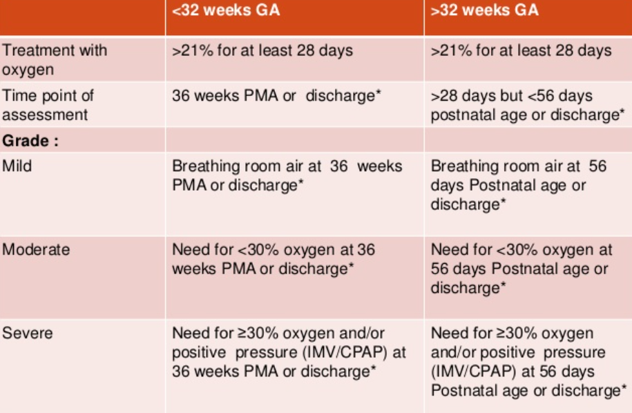
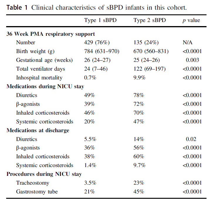
Not all BPD is the same though. in 2000 the NIH produced a new consensus definition of BPD as shown below.
What stands out for the babies <32 weeks is how severe BPD is defined. Babies who are ventilated are classified in the same severity group as those who are on CPAP. Somehow that doesn’t seem quite right intuitively but alas that is what they decided at the time.
Type 1 sBPD: patients on nasal cannula or noninvasive positive pressure support (i.e., high flow nasal cannula (HFNC), nasal continuous positive airway pressure (nCPAP), noninvasive intermittent positive pressure ventilation (nIPPV)) Type 2 sBPD: infants receiving iMV
The authors then looked at a sample of 564 patients from 2015-2019 in the BPD collaborative registry and subdivided them into 429 (76%) Type 1 vs 135 (24%) Type 2 sBPD and compared outcomes between the two. The differences between the two types of BPD are quite significant and shown in Table I. Babies who went on to develop sBPD as Type 2 were younger and smaller than those with Type 1. Medication use within the NICU and after discharge was markedly different as were the total ventilator days which is likely not surprising since by definition they were still intubated at 36 weeks. Importantly if you were still intubated at 36 weeks PMA almost one quarter of the patients went on to receive a tracheostomy.
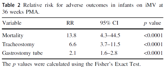
Looking at it another way using relative risks the signifance of having Type 2 sBPD is impactful.
Taking Meaning From This
You might be quick to say, Michael this is absolutely no surprise. On the other hand if you have read this blog for some time you may remember this piece The New BPD That Matters. This study looked at what gestational age really mattered when looking at long term pulmonary outcomes in a Canadian cohort. When you take all comers it was 40 weeks and not 36 weeks that really mattered. The likely differernce here though is that by selecting out only the severe patients in this current study it is indeed the 36 week mark that still has relevance. I actually think the two papers together are not contradictory but rather additive.
What I think one takes away from the current study is that failure to extubate by 36 weeks does in fact carry with it significant long term risk to the patient. It would be easy enough to say that these babies should be extubated but as you see from table I it isn’t that they didn’t try. From a medication standpoint it would appear that they ” threw the kitchen sink” at these babies. The only thing I find a little surprising is that only 47% of babies in the collaborative with type 2 sBPD received systemic steroids. If they were that sick I would have expected it to be higher although that also may just be a reflection of my own practice.
One thing that I think will be a hot topic moving forward is the use of higher levels of CPAP than what many units are accustomed to. This has also been recently discussed in High CPAP vs NIPPV. Is there a winner? There may be a reluctance by some units to use CPAP levels in the +9-12 cm H2O range but when looking at these downstream complications for patients who remain ventilated at 36 weeks I think people need to seriously consider their biases and whether they are based on science or what they were taught. I can’t help but think of the oft used expression absence of evidence is not evidence of absence and think that if we can all be a little humble who knows what we may discover that can help this population.
Who doesn’t love a good match up?! Supporting neonates in need of resuscitation after delivery has been the subject of many studies over the years. The movement has certainly been to non-invasive support with CPAP or NIPPV but some babies need some degree of support with PPV after delivery when they simply won’t breathe. Prior to intubation the rise of the t-piece resuscitator has meant that practitioners can set a PIP and PEEP and with only a finger press to deliver a tidal volume at set pressure and with the finger released provide CPAP through the same device. The only problem potentially with use of these devices is the imposed work of breathing (iWOB) which has been measured in other studies. Any device I have used has provided ventilation through a mask so imagine my surprise to come across a new device called rPAP using prongs from the original infant flow design. From the manufacturers website the company claims that their design used with either a mask or nasal prongs reduces iWOB by 92% compared to other comparable machines! Imagine my greater surprise to see a head to head RCT comparing this new device to standard t-piece resuscitators with a mask.
The intervention was completed with one of three outcomes were met.
Infant intubated
Stable and breathing on method of support after a minimum of 10 minutes of support.
At 30 minutes when respiratory support could continue as decided by the clinician without crossover allowed.
Looking at the appendices for the trial it appears that one could use either device to administer PPV or CPAP but the point of the trial was that the devices would be used to support the infants until one of the three above criteria were met. If the claims about reduced iWOB were true compared to other devices in use then one might expect to see a difference in the primary outcome of incidence of intubation or death within 30 minutes of birth.
In total there were 250 infants recruited with 127 assigned to the rPAP and the other 123 to t-piece resuscitation. The mean GA in the trial was 24.8 weeks and the baseline characteristics between groups were similar although the group randomized to the rPAP has more c-sections and more general anesthetic exposure compared to the t-piece group. Lastly, humidification of gases during resuscitation was similar between the two groups.
How Did They Compare?
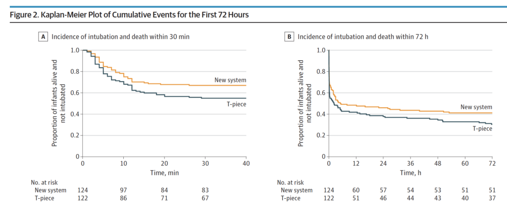
It just might be that the claims of decreased iWOB might have merit. In Figure 2 below the Kaplan-Meier curves show a difference favouring the rPAP device when looking at the primary outcome. This difference was significant with 41 of 124 infants (33.1%) in the rPAP group and in 55 of 122 infants (45.1%) in the T-piece group having the primary outcome of intubation or death within the first 30 minutes of life. Moreover when looking at the adjusted odds ratio it was still significant at 0.53; 95% CI, 0.30-0.94. The incidence of intubation and death in the first 72 hours although trending towards favouring the new system did not reach statistical significance.
Finally, none of the secondary outcomes reached statistical significance which included such things as death in the delivery room, use of surfactant, or PPV in the DR.
Does it make sense?
If you had asked me to tell you prior to the study whether resuscitation with nasal prongs vs a mask would be different I would have said a mask would be better due to less leak. Turns out based on this data that I would be wrong in that guess. A look at the website though for the rPAP device indicates that it can be used with a mask or nasal prongs. It would have been nice in the study presented here to have used a mask as a third arm with the rPAP device as it leaves me wondering a bit whether it was the interface that mattered more than the type of driver used? Maybe I am wrong and by using prongs it allows the infant to have less iWOB than with a mask over the mouth and nose? Could it be that it has more to do with that that the type of driver whether it is a traditional t-piece resuscitator or the new rPAP device? Regardless, I have a suspicion that these results will resonate with people. A posting of the abstract alone has garnered a lot of attention on twitter this week so clearly this is of interest.
I don’t think there is much fault to find in this study other than my question of why they didn’t choose to have a head to head comparison with masks as well but perhaps that is for another study. I imagine we will see this approach adopted in many centres around the world as they replace their traditional t-piece resuscitators in need of replacement. I also suspect there will be many that will want a larger study before adopting this strategy to look more closely with come faith in the results at secondary outcomes in particular having to do with safety.
One thing is for certain. There will be more studies to come!
Here in Winnipeg we don’t use t-piece resuscitators for any resuscitation. I did use them in my past position in Edmonton and I came to appreciate them for their ease of use. For the majority of infants, setting a PIP and a PEEP and then using your finger to occlude and release offers a relatively simple and less difficult approach to ventilation than using a self inflating or jackson-rees bag. I say the majority of infants, as most infants are not born from 22-32 weeks but the lion’s share are born at gestations older than that. The larger more mature infants have lungs that are much more forgiving to excessive ventilation. For the smallest of infants though questions have remained for some time around the volumes delivered to the fragile lung when a fixed pressure is used in the presence of moment to moment changes in compliance.
Measuring Tidal Volume in Intubated At Risk Infants
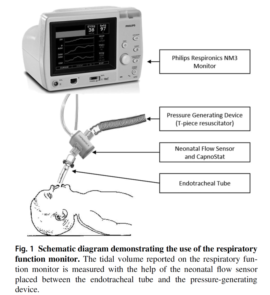
Vaidya R et al published Tidal volume measurements in the delivery room in preterm infants requiring positive pressure ventilation via endotracheal tube feasibility study in Journal of Perinatology. The prospective observational study looked at 10 infants born at < 32 weeks with a mean GA of 23.9(±1.5) weeks and mean BW 618.5(±155)g. A mean of 17.8 minutes of recordings were examined using the setup below and in total looked at 8175 individual breaths. All patients in the study were intubated with non-cuffed ETT but by only including intubated infants in the delivery room the issue of mask leak was avoided. As in many units the target Vt was 4-6 mL/kg. It wasn’t specified what criteria they use for setting initial pressures but the included patients had a mean PIP of 24.4±5 and PEEP of 5.9 ±2.4. Importantly, those providing ventilation with the t-piece resuscitator were blinded to the data on tidal volume measurements.
How Good Were They At Meeting Their Goal?
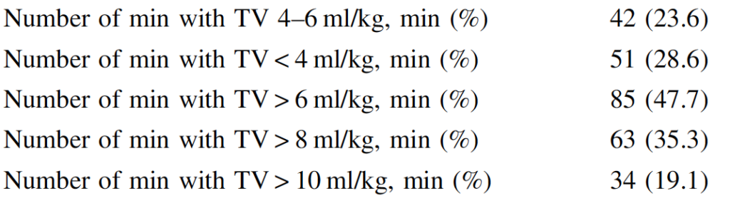
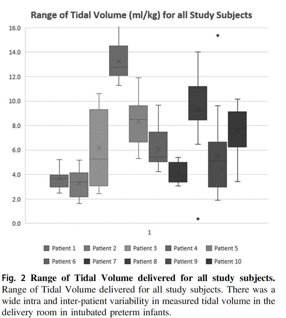
It turns out that they weren’t that great (I am not faulting them by the way) as it is a challenge to try and adjust pressures based on chest rise. We are not good at it at all. As shown in the figure below there was a wide range of volumes administered. In fact here is the breakdown. The goal Vt between 4-6 was only 25% of the time. In other words you are dealing with either a risk of atelectotrauma or volutrauma 75% of the time. It is worth noting that the neonatal flow sensor has a dead space of 1 mL. If that is the case and the infants on average were about 600g that is almost 2 mL/kg in non-ventilated space that this volume is going into. It doesn’t change the numbers that much if you factor that in but it does mean that some infants who were getting a measured 3 mL/kg were actually seeing under 2 mL/kg of lung ventilation. On the other hand those getting 7 mL/kg were actually seeing under 6 mL/kg so were in target. Bottom line though is that when using fixed pressure settings in the presence of changing compliance even if one is adjusting pressure in real time it is difficult to maintain stable volumes in target range. The authors also demonstrate in another graph that even in individual patients there is fluctuation as well.
Call to Action
I think this study is actually quite useful in confirming what I imagine many have always suspected. We just aren’t that great at assessing tidal volume when we watch the chest rise. As many have noted, the first 6 breaths at least in an animal model can damage the lungs. Imagine what excessive or low volumes can do to the lung over 18 minutes?!
What this study does is demonstrate especially in the smallest and most vulnerable infants that if ventilation is needed one should put the infant onto a volume guaranteed mode of ventilation ASAP. Ventilators should be in the resuscitation area as we have in our hospital and not have to be brought in should the baby be intubated. Hand bagging even with a t-piece resuscitator should be kept to a minimum. At risk is the development of BPD and knowing that even in experienced hands we just aren’t that good at delivering tidal volumes in a target range we need to strive to minimize the time that we expose our infants to such modalities. Ventilation isn’t always avoidable but when needed my advice is to control volume and allow pressures to fluctuate as resistance and compliance change. Especially after administration of surfactant the pulmonary mechanics are changing constantly and no matter how good you are you just won’t be able to keep pace. Let the ventilator do it!